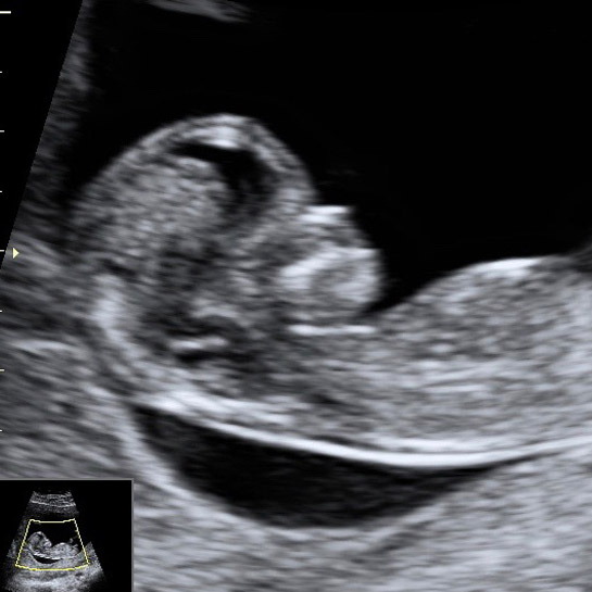
Dating nuchal thickness
Contents:
A nuchal translucency scan is part of the screening tests for Down Syndrome. What is a nuchal translucency scan? What does a nuchal translucency scan look for? How is a nuchal translucency scan performed and when? What happens if my baby is in the high risk category?
- Optimising the timing for nuchal translucency measurement.;
- faisalabad dating sites?
- carbon dating prices?
- What is a nuchal translucency scan?.
- week pregnancy dating scan - NHS!
- What is the purpose of the dating scan?.
Do I have to have the test? No, it is your decision. Or you might decide not to have the nuchal translucency scan, or any other tests. Opens in a new window.
Department of Health Guidelines for the use of ultrasound in the management of obstetric conditions. Raising Children Network How to decide on antenatal tests for chromosomal abnormalities and other conditions. Raising Children Network Antenatal tests: Was this article helpful? Nuchal Translucency Scan - InsideRadiology. Pregnancy - prenatal tests - Better Health Channel. Find out about early ultrasounds at weeks, sometimes called dating scans. Screening for Down syndrome. Checkups, scans and tests during pregnancy. Questions to ask your doctor about tests and scans. Sorry, no results were found for ""Nuchal translucency scan"".
There was an error contacting server. These first-trimester serum markers are described independent of NT, which would imply that a unified protocol using both serum markers and ultrasound can be applied with more accuracy than either alone. Nuchal translucency and first and second trimester serum markers; Integrated testing. This two-step testing involves a combination of NT and pregnancy-associated plasma protein A in the first trimester with serum AFP, hCG, unconjugated E3, and Inhibin-A in the second, with a single DS result being provided in the second trimester.
This factor may lead to unnecessary anxiety for the patient and concerned family. Nasal translucency and absence of nasal bone in first-trimester ultrasound. The relationship of increased NT and absence of fetal nasal bone has been coined as an ultrasonic screening tool during the first-trimester but adequate visualization of the nasal bone needs expertise and correct technique. This report was challenged by Hutchon et al. More evidence-based studies are needed to validate the importance of absent nasal bone as a screening marker for DS. First trimester screening holds the promise of improved detection rates with lower false-positive rates.
INTRODUCTION
The emerging effects and possible pathogenic mechanisms of enlarged NT include fetal heart failure secondary to a cardiac defect, anemia, infection, inappropriate expression of atrial natriuretic peptide; abnormal extracellular matrix; or abnormalities of lymphatic structure and drainage [ 40 ]. Enlarged NT leads to lymphatic obstruction which in its most severe form results in cystic hygroma.
- interactive dating sims?
- 10 year old dating website?
- online dating farmers nz?
- Nuchal scan;
- Page contents.
- The Associations of Nuchal Translucency and Fetal Abnormalities; Significance and Implications;
A cystic hygroma is a fluid-filled multi-septated cyst or cysts that arise from the back of the neck. When an enlarged NT or small cystic hygroma resolves before birth, the infant may be left with a webbed neck. Clark [ 41 ] and Lacro [ 42 ] reported a strong association between webbed neck and coarctation of the aorta in infants with Turner syndrome. The reported association between NT-webbed neck and cardiac anomalies, both in fetuses with a variety of genetic syndromes and in euploid fetuses, points to the possibility of an established relationship.
The lymphatic obstruction that leads to an enlarged jugular lymph sac could also cause lymph to accumulate in the thoracic duct. Due to its anatomical location, in the thoracic cavity, the enlarged thoracic duct might exert pressure on or displace the heart, causing obstruction of blood flow through the cardiac chambers, and leading to abnormal inadequate growth of certain cardiac structures.
A nuchal translucency scan (NT scan) is an ultrasound screening test for assessing So the NT scan will usually happen alongside your routine dating scan. A nuchal scan or nuchal translucency (NT) scan/procedure is a sonographic prenatal screening The scan may also help confirm both the accuracy of the pregnancy dates and the fetal viability. As nuchal translucency size increases, the.
Cardiac anomalies believed to result from such abnormal intracardiac blood flow include aortic coarctation and hypoplastic left heart [ 43 ]. Currently, screening by fetal echocardiography is offered to the fetus following the observation of an NT of 3. The cost-effectiveness of offering fetal screening echocardiography at NT measurements of 2.
Hyett and coworkers [ 45 ] evaluated the relationship between NT size and major cardiac defects in more than 29, euploid pregnancies, and described that the incidence of cardiac defects increased along with NT size; the prevalence was only 0. Goetzl [ 46 ] has summarized results from seven studies that the incidence of cardiac abnormalities was positively related to NT; for NT of up to 3. There is growing body of evidence that patients with increased fetal NT and normal karyotype are at higher risk of adverse outcome, cardiac or otherwise [ 10 ].
Cardiovascular anomalies are the most frequently encountered defects in chromosomally normal fetuses with increased NT. Based on such findings, early fetal echocardiography and anomaly scan should be considered in these fetuses. Enlarged NT has been reported with other structural anomalies, including diaphragmatic hernia, exompholos, body stalk anomaly, fetal akinesia syndrome, skeletal dysplasias, various multiple anomaly syndromes, and fetal loss [ 47 , 48 ].
The first-trimester maternal serum screening has consistently revealed that pregnancies with fetal DS are associated with higher levels of total hCG and of the free hCG with a median multiple of the median [MoM] of 1. The actual anatomic structure whose fluid is seen as translucency is likely the normal skin at the back of the neck, which either may become edematous or in some cases filled with fluid by dilated lymphatic sacs due to altered normal embryological connections. Prenatal Ultrasound is a widely accepted tool for detecting fetal anomalies during pregnancy and, once detected, further investigations are instigated, including fetal chromosome analysis, maternal and fetal investigations for infections, microarray analysis, and fetal echocardiogram and magnetic resonance imaging, when indicated. A normal 20 week scan of a euploid fetus with a history of first trimester increased nuchal translucency: Checkups, scans and tests during pregnancy. You can ask your midwife or doctor before the scan if this is the case.
Keeping in view the published literature about the associations of enlarged NT, the euploid should also be evaluated by targeted second trimester ultrasound examination [ 49 ]. An increased NT has been associated with parvovirus infection [ 50 ].
We value your feedback
If increased NT leads to signs of fetal hydrops at 20 to 22 weeks, parvovirus screening is recommended, in addition to evaluating the standard infections associated with fetal hydrops, such as toxoplasmosis and cytomegalovirus [ 46 ]. Associations of increased NT have also been described with cerebral hypoplasia [ 51 ], facial cleft [ 52 ], spine disorganization [ 53 ], hydrops and hepatomegaly [ 54 ], growth retardation [ 55 ], and skin edema [ 56 ].
Ultrasound examination of the fetus is a subjective process that is highly dependent on operator skills and the quality of the sonographic equipment. These limitations militate against the deployment of ultrasound as a screening tool in the manner in which maternal serum biochemistry has been used [ 57 ]. Evaluation of the nuchal translucency should be considered during the first trimester ultrasound and a detailed anatomic evaluation should be offered whenever feasible.
Increased NT is associated with a spectrum of fetal abnormalities.
Your pregnancy and baby guide
The commonest association is with chromosomal defects. In fetuses with increased NT and a normal karyotype, the risk of an adverse outcome remains and increases with increasing NT. An NT of 3. Patients should be counseled for increased risk of fetal loss before embarking upon any invasive manipulation. Fetal outcome is favorable in the absence of any identified abnormalities and with resolution of NT thickening in the progressive scans. For reproducible and accurate measurements of NT, strict adherence to quality guidelines of the technique, training and supervision of the sonologist is of utmost importance.
National Center for Biotechnology Information , U. J Clin Diagn Res. Published online Mar Shaista Salman Guraya 1. Find articles by Shaista Salman Guraya.

Author information Article notes Copyright and License information Disclaimer. This article has been cited by other articles in PMC. Abstract This review of literature describes the first-trimester nuchal translucency NT which forms the basis of new form of screening which can lead to a significant improvement in detection of congenital anomalies as compared to second trimester screening programs, the so called genetic-sonogram.
Open in a separate window. Sonographic criteria to maximize quality of nuchal translucency sonography. Nuchal translucency ultrasound should only be performed by sonog raphers or sonologists trained and experienced in the technique. Transabdominal or transvaginal approach should be performed, based on maternal body habitus, gestational age, and fetal position. Gestation should be limited to between 10 weeks 3 days and 13 weeks 6 days approximate fetal crown—rump length, 36—80 mm. At least three nuchal translucency measurements should be obtained, with the mean value of those used in risk assessment and patient counseling.
The scan may also help confirm both the accuracy of the pregnancy dates and the fetal viability. All women, whatever their age, have a small risk of delivering a baby with a physical or cognitive disability.
The Associations of Nuchal Translucency and Fetal Abnormalities; Significance and Implications
The nuchal scan helps physicians estimate the risk of the fetus having Down syndrome or other abnormalities more accurately than by maternal age alone. Overall, the most common chromosomal disorder is Down syndrome trisomy The risk rises with maternal age from 1 in pregnancies below age 25, to 1 in at age 35, to 1 in at age In , Sequenom announced the launch of MaterniT21, a non-invasive blood test with a high level of accuracy in detecting Down syndrome and a handful of other chromosomal abnormalities. As of , there are five commercial versions of this screen called cell-free fetal DNA screening available in the United States.
Blood testing is also used to look for abnormal levels of alphafetoprotein or hormones. The results of all three factors may indicate a higher risk.
If this is the case, the woman may be advised to have a more reliable screen such as cell-free fetal DNA screening or an invasive diagnostic test such as chorionic villus sampling or amniocentesis. Screening for Down syndrome by a combination of maternal age and thickness of nuchal translucency in the fetus at 11—14 weeks of gestation was introduced in the s. In fetuses with a normal number of chromosomes, a thicker nuchal translucency is associated with other fetal defects and genetic syndromes.
Nuchal scan NT procedure is performed between 11 and 14 weeks of gestation, because the accuracy is best in this period. The scan is obtained with the fetus in sagittal section and a neutral position of the fetal head neither hyperflexed nor extended, either of which can influence the nuchal translucency thickness.
It is important to distinguish the nuchal lucency from the underlying amniotic membrane. Normal thickness depends on the crown-rump length CRL of the fetus. Among those fetuses whose nuchal translucency exceeds the normal values, there is a relatively high risk of significant abnormality. Further, other, non-trisomic abnormalities may also demonstrate an enlarged nuchal transparency. This leaves the measurement of nuchal transparency as a potentially useful first trimester screening tool. Abnormal findings allow for early careful evaluation of chromosomes and possible structural defects on a targeted basis.
How to define a normal or abnormal nuchal translucency measurement can be difficult.